小脑齿状核钙化图片

萌妹子课堂——颅内钙化知多少(生理篇)
图片尺寸640x723
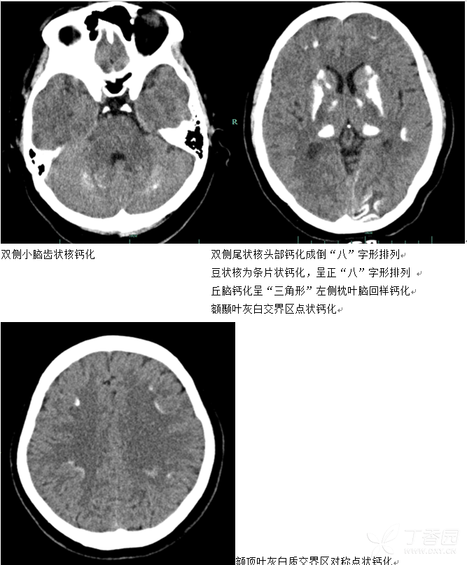
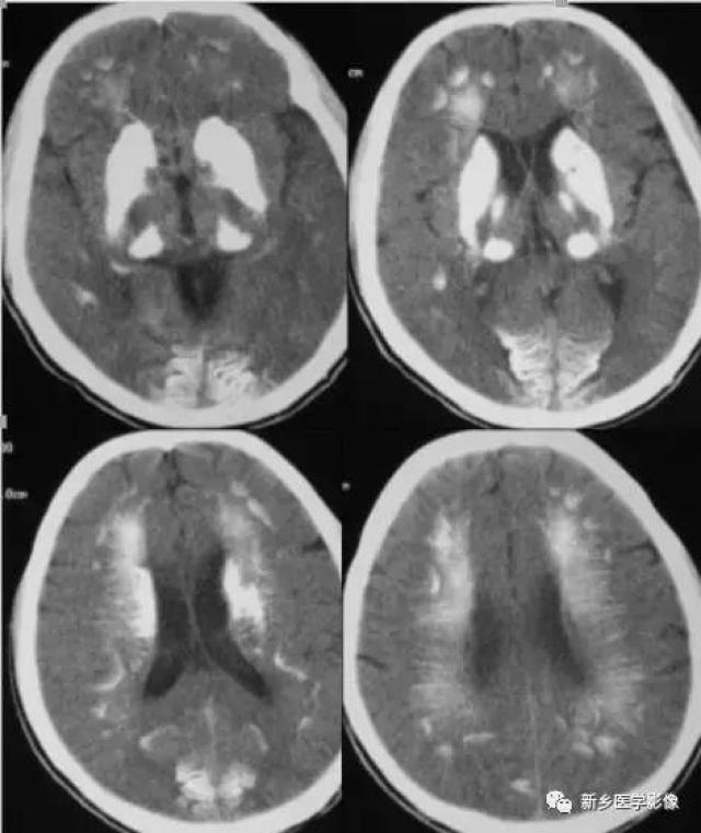
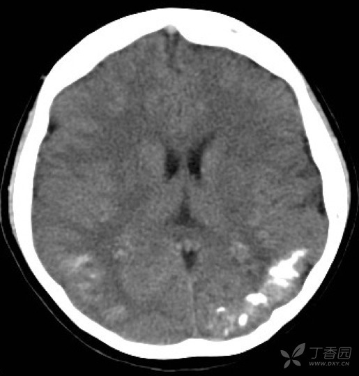
头颅ct(见下图):双侧小脑,基底节区,灰白质交界区广泛对称性钙化以及
图片尺寸667x807
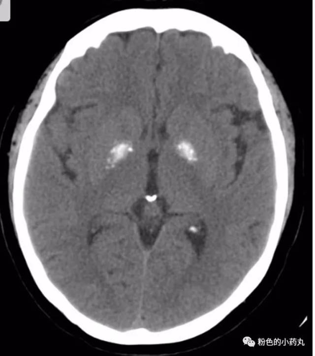
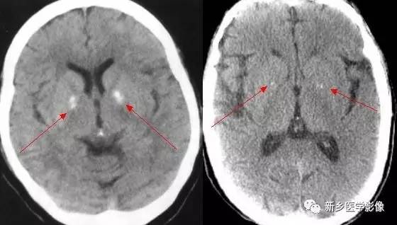
状核,尾状核,丘脑及双侧灰白质交界带对称性斑片状,条状高密度钙化影
图片尺寸450x600
图解颅内出现这7个部位的钙化属正常
图片尺寸556x604
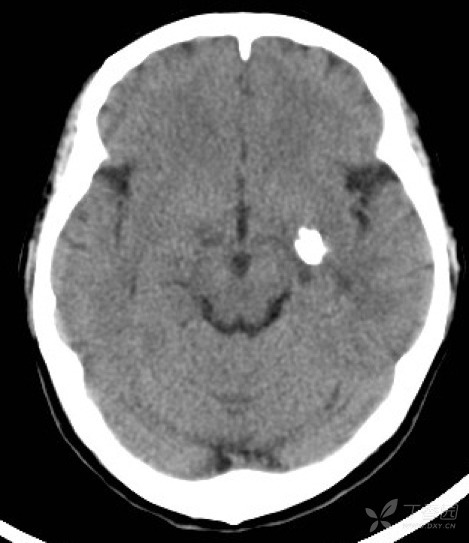
简要病史:男性,89 岁,体检小脑齿状核钙化基底节钙化的基本化学成份与
图片尺寸1015x512
小脑钙化
图片尺寸640x294
常见颅内钙化的影像学征象
图片尺寸640x412
这样的颅内钙化见一次就终身难忘
图片尺寸596x399
31岁男子频繁呕吐1个月头晕1周罪魁祸首竟然是
图片尺寸636x707
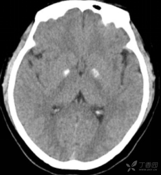
小脑齿状核钙化相对少见,表现为对称性高密度钙化.生理性病理性均可.
图片尺寸560x318
干货!颅脑 ct 钙化模式对疾病鉴别诊断意义
图片尺寸775x492
核形钙化:钙化与尾状核,豆状核,丘脑,小脑齿状核形态一致.
图片尺寸800x800
图解颅内出现这7个部位的钙化属正常
图片尺寸469x543
34种颅内可以钙化的病变汇总
图片尺寸472x561
看了那么多颅内钙化,这些鉴别「套路」你可知晓?
图片尺寸832x624
图解颅内出现这7个部位的钙化属正常
图片尺寸515x539
2002年的ct片是双侧苍白球钙化,2009年发展为双侧小脑钙化
图片尺寸480x360
常见颅内钙化的影像学征象
图片尺寸958x569
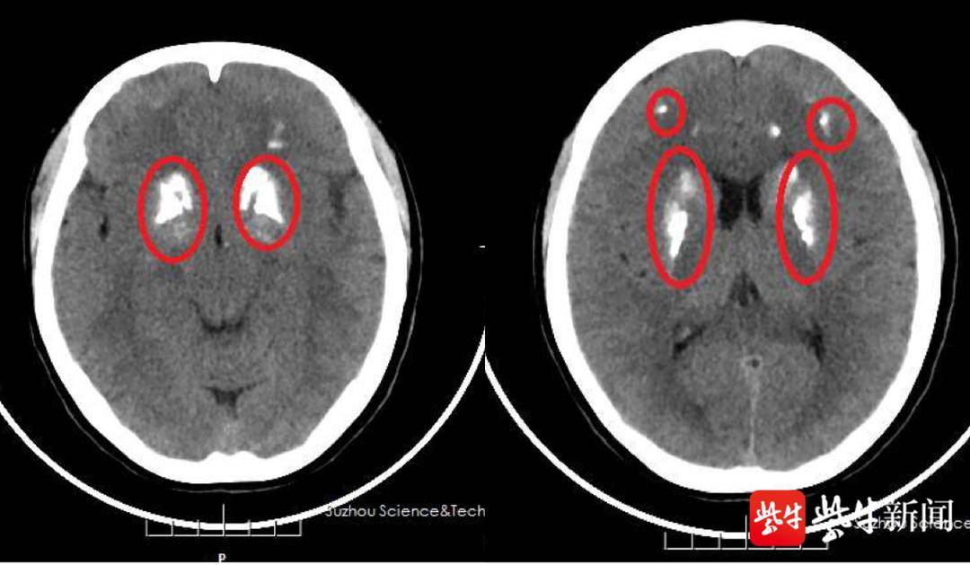
双侧基底节钙化,如何诊断?
图片尺寸507x618
两侧大脑及小脑出现多发对称性钙化影
图片尺寸1080x629